Morning Blur That Clears
Blurry or foggy vision on waking that improves later in the day — one of the earliest and most telling signs.


Fuchs’ dystrophy is a hereditary condition that gradually destroys the cells your cornea needs to stay clear. Left untreated it leads to permanent vision loss, but caught early it can be managed — and when surgery is needed, DMEK offers excellent outcomes with low rejection risk and fast recovery. Diagnosis, medical management, and DMEK surgery by cornea specialist Dr. Bonnie Sklar.

 Clinical image. Individual presentation varies.
The cornea — the clear front surface of your eye — depends on a single layer of cells on its inner surface to stay transparent. These corneal endothelial cells act as pumps that continuously remove excess fluid. When they work properly the cornea stays clear; when they don't, it swells with fluid and clouds your vision.
In Fuchs’ endothelial dystrophy, a hereditary condition, these cells gradually deteriorate and die. As the cell count drops, the cornea loses its ability to regulate fluid and begins to swell — a process called corneal edema — producing increasingly blurry, foggy vision that worsens over time. It affects roughly 4% of people over 40, is more common in women, and often isn't noticed until vision changes appear in a person's 50s or 60s. A telling early sign is morning blur that clears as the day goes on, because the closed eye can't evaporate excess fluid overnight.
See the StagesSymptoms usually begin gradually and worsen over years. Early disease may cause only mild disturbances; advanced disease significantly impairs vision.
Blurry or foggy vision on waking that improves later in the day — one of the earliest and most telling signs.
Glare and halos around lights, particularly at night or in bright conditions, with difficulty driving at night due to reduced contrast.
Increased sensitivity to light (photophobia) as the disease progresses.
Vision that looks cloudy or washed out regardless of your glasses prescription.
Fluctuating vision that your optometrist cannot fully correct with a new prescription.
In advanced disease, persistent blur throughout the day and eye pain from surface blistering (bullous keratopathy).
If Fuchs’ runs in your family: regular corneal evaluations are recommended even before symptoms appear. Early detection significantly expands your treatment options and timeline.
Fuchs’ progresses through distinct stages over years or decades. Understanding your current stage is essential to determining the right treatment approach — and the right timing.
Tiny deposits called guttae appear on the corneal endothelium and cell density begins to decline. Vision may be normal or only mildly affected, and most patients are unaware they have Fuchs’. Management focuses on monitoring, with hypertonic saline drops to ease morning blur.
The cornea begins retaining fluid in the stroma. Vision becomes noticeably blurry, especially in the morning, and glare and halos worsen. Corneal thickness increases measurably on pachymetry. Hypertonic drops and the hair-dryer technique provide temporary relief, and surgical planning often begins here.
Fluid breaks through to the outer surface, forming painful blisters called bullae. Vision is severely impaired throughout the day and scarring may begin to develop. A corneal transplant — specifically DMEK — is typically required to restore vision and relieve pain.
Treatment depends entirely on the stage of the disease. Early Fuchs’ is managed medically; advanced disease is treated surgically — and today's options are dramatically better than a decade ago.
Early to moderate disease. Sodium chloride drops (5% NaCl) draw excess fluid out of the swollen cornea by osmosis. They don't stop the disease but can significantly reduce morning blur and improve daily comfort. Dr. Sklar recommends the appropriate formulation and frequency for your severity.
Practical management. Holding a hair dryer at arm's length and directing warm (not hot) air toward your face on waking helps evaporate surface moisture, easing morning blur. This is a genuine clinical recommendation, and combined with hypertonic drops it makes early Fuchs’ much more manageable.
Advanced disease — first-line surgery. Descemet Membrane Endothelial Keratoplasty replaces only the diseased Descemet membrane and endothelial cells with a thin donor graft, typically giving excellent visual outcomes, fast recovery, and the lowest rejection rate of any corneal transplant. Learn about DMEK.
Alternative surgical option. When DMEK isn't the best fit, DSEK replaces the inner corneal layers with a slightly thicker donor graft. It remains an excellent option with far faster recovery than full-thickness transplantation. Compare transplant types.
Lowest
Rejection risk of any corneal transplant
Weeks
Typical onset of visual improvement
~3mm
Incision size — no large corneal wound
No
Corneal graft sutures required
Why DMEK is preferred for Fuchs’: because only the diseased inner layer is replaced — not the entire cornea — DMEK is today's standard of care, with recovery, rejection risk, and visual outcomes that are significantly better than older full-thickness PKP transplantation.
Diagnosing Fuchs’ and staging it accurately requires more than a standard eye exam. Dr. Sklar combines advanced imaging and clinical tests to build a complete picture of your corneal health.
The primary tool for detecting guttae, the tiny corneal deposits that are the hallmark of Fuchs’. A trained cornea specialist can detect early guttae before any symptoms occur.
Photographs and counts the corneal endothelial cells for a precise density measurement. Declining cell counts are a key marker of progression and help guide the timing of surgery.
Measures corneal thickness at multiple points. A cornea swelling with fluid is measurably thicker than normal, and pachymetry quantifies this and tracks change over time.
3D mapping of the corneal surface and internal structure, used to detect irregular astigmatism from swelling and to guide surgical planning for DMEK.
Dr. Sklar is Berg-Feinfield's fellowship-trained cornea specialist, with particular expertise in the surgical management of Fuchs’ endothelial dystrophy.
Completed her cornea fellowship at Duke University Eye Center, one of the nation's most respected programs, and her residency at Wills Eye Hospital in Philadelphia.
Her practice centers on the full range of corneal transplantation — DMEK, DSEK, and PKP — along with keratoconus, cross-linking, and ocular surface disease.
For patients considering DMEK, her fellowship training provides the specialized background this technically demanding procedure requires.
You don't need to be symptomatic to benefit — early staging establishes a baseline and allows for properly timed intervention if the disease progresses.
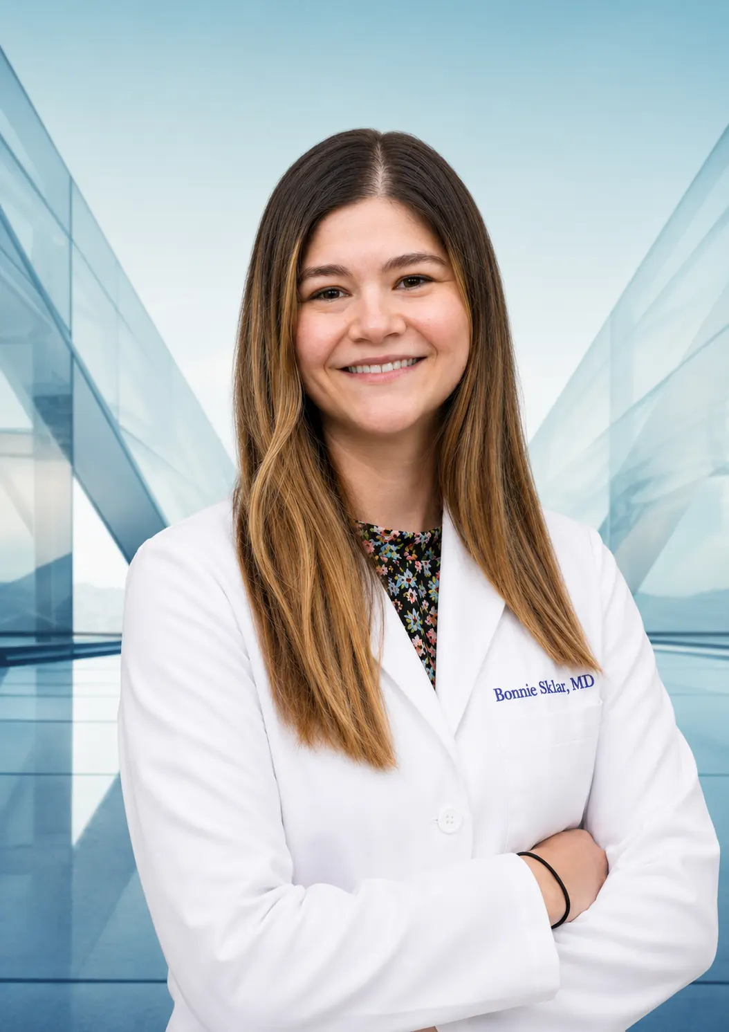
Dr. Sklar sees Fuchs’ dystrophy patients across Berg-Feinfield's Los Angeles-area offices, with corneal evaluations and DMEK surgical consultations available throughout the region.
2625 W. Alameda Ave.
Suite 208
Burbank, CA 91505
(818) 845-3557
13320 Riverside Drive
Suite 114
Sherman Oaks, CA 91423
(818) 501-3937
462 N. Linden Drive
Suite 441
Beverly Hills, CA 90212
(866) 273-3327
638 W. Duarte Road
Suite 10
Arcadia, CA 91007
(626) 795-9793
27335 Tourney Road
Suite 210
Valencia, CA 91355
(866) 273-3327
Whether you've just been told you have guttae or you've been living with Fuchs’ dystrophy for years, Dr. Sklar and the Berg-Feinfield cornea team will give you an honest assessment of where you are and what your options are.
BERG-FEINFIELD · AFTER HOURS
Questions about your vision? Talk with our AI assistant about LASIK, cataract surgery, or general eye care.
Talk with ClaireAI assistant for general information, not medical advice or emergency care. AI can make mistakes. Confirm medical information with your eye care professional.