A Visible Growth
A pinkish or whitish growth on the white of the eye, usually on the side closest to the nose.

Pterygium — commonly called surfer's eye — is one of the most common eye conditions in sunny, outdoor-active cities like Los Angeles. It's treatable, and when surgery is needed, the right technique makes all the difference in preventing it from coming back. Diagnosis, management, and surgical removal by fellowship-trained cornea specialist Dr. Bonnie Sklar.
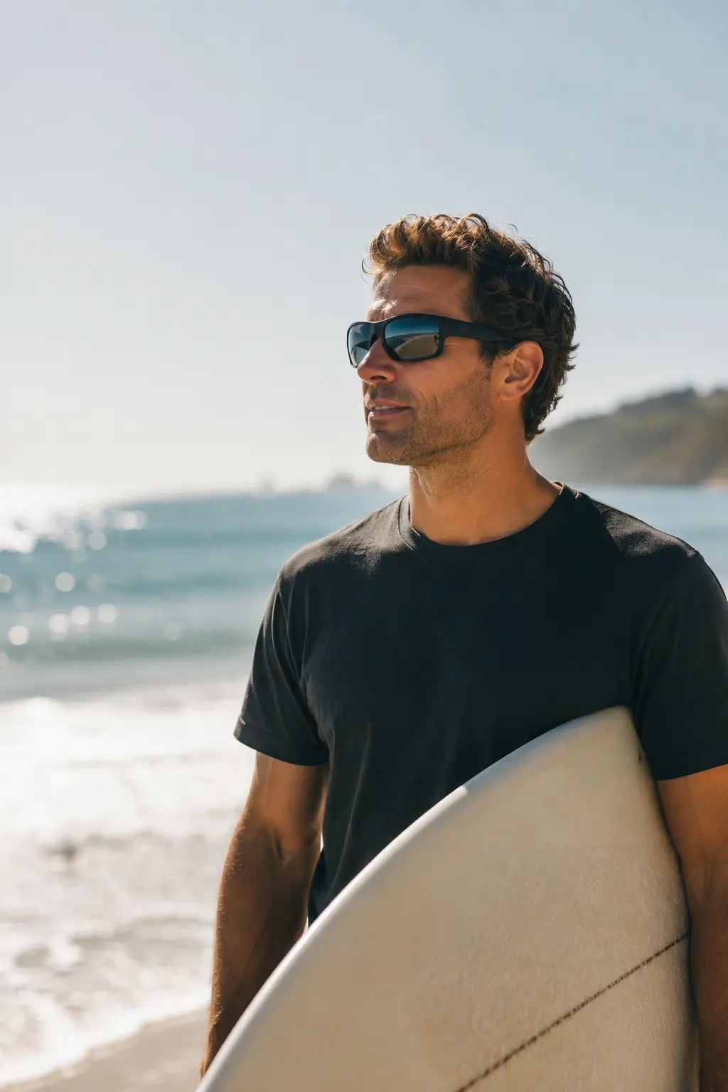
 Clinical photograph. Individual presentation varies.
A pterygium (pronounced teh-RIJ-ee-um) is a non-cancerous growth of fibrovascular tissue that begins on the conjunctiva — the clear membrane covering the white of your eye — and spreads onto the surface of the cornea. It typically starts on the nasal side and grows horizontally toward the pupil, and in some cases both eyes are affected.
The tissue is wedge-shaped and may look pink, fleshy, or slightly opaque. Small pterygia often cause nothing more than mild redness and irritation, while larger ones can induce astigmatism, distort vision, and in advanced cases cover the visual axis. A pterygium is not cancerous and won't spread through the body, but it won't go away on its own — once it grows toward the center of the cornea, monitoring and eventual removal are typically recommended.
See the GradesSymptoms range from mildly bothersome to vision-impairing, depending on the size and location of the growth.
A pinkish or whitish growth on the white of the eye, usually on the side closest to the nose.
Persistent redness or inflammation in the affected area, sometimes with sensitivity to light, wind, or dust.
A gritty feeling, as though something is in your eye.
Burning, stinging, or excessive tearing associated with the growth.
Particularly if the pterygium is inducing astigmatism as it grows onto the cornea.
The growth is visible and can be socially noticeable, which prompts some patients to seek treatment.
When to see a specialist: if your pterygium is growing, causing significant discomfort, distorting vision, or approaching the pupil, see a cornea specialist. The longer it grows onto the cornea, the more tissue is involved and the more complex surgery becomes — earlier is better.
Pterygia are graded by how far they extend onto the cornea. Your grade determines the urgency of treatment and influences the surgical approach if removal is needed.
The pterygium reaches the edge of the cornea (the limbus) but has not grown onto the corneal surface. Little or no corneal involvement, and usually no symptoms beyond mild redness.
The pterygium extends onto the cornea but has not reached the pupil margin. It may cause mild astigmatism and a foreign-body sensation. Managed conservatively or surgically depending on growth rate.
The pterygium reaches the edge of the pupil, and significant induced astigmatism and visual distortion are common. Surgical removal is typically recommended at this stage.
The pterygium crosses the pupil and obscures the visual axis, causing significant vision loss. Removal is urgent, though corneal scarring beneath the growth may affect the final visual outcome.
Treatment depends on grade, growth rate, and symptoms. Many small pterygia are monitored for years; when growth, vision impact, or comfort cross a threshold, surgery is the only definitive treatment.
Artificial tears reduce dryness, irritation, and the foreign-body sensation. They don't stop growth but improve comfort, and are appropriate for mild, non-progressive pterygia.
Wraparound UV400 sunglasses, wide-brimmed hats, and reducing peak-hour sun exposure are the most important steps for slowing growth and preventing recurrence — especially in year-round-sunny Los Angeles.
When a pterygium becomes acutely inflamed, a short course of mild topical steroid or non-steroidal anti-inflammatory drops can calm it. This is a temporary measure and does not treat the underlying growth.
The only way to remove a pterygium permanently. Dr. Sklar uses the conjunctival autograft technique — the current gold standard — which has a significantly lower recurrence rate than older approaches.
The pterygium is removed and the bare area is replaced with a small graft of the patient's own healthy conjunctiva — typically taken from the upper part of the same eye, hidden under the eyelid.
The abnormal fibrovascular tissue is carefully excised from the corneal surface and surrounding conjunctiva under topical anesthesia.
Any residual tissue is removed and the underlying corneal surface is prepared to minimize scarring and promote clear healing.
A thin piece of healthy conjunctival tissue is taken from the upper portion of the same eye, an area that heals without visible consequence.
The graft is placed over the bare area and secured with fibrin glue or fine sutures, acting as a barrier to prevent regrowth of fibrovascular tissue.
Most patients return to normal activities within one to two weeks. The eye may be red and mildly uncomfortable for several days, and anti-inflammatory drops are used during healing.
On recurrence: no technique eliminates it entirely, but the conjunctival autograft significantly reduces the risk compared to bare-sclera excision. Diligent UV protection after surgery is the single most important thing a patient can do to keep a pterygium from coming back.

Pterygium is largely a UV-related condition, and in Los Angeles year-round sun and an outdoor lifestyle add up quickly.
Cumulative ultraviolet exposure — not a single sunburn, but decades of surfing, hiking, cycling, commuting, and working outdoors — is what drives the fibrovascular tissue to grow. Wind and dust accelerate it, and Santa Ana conditions deliver both. The habits below meaningfully reduce both first-time occurrence and post-surgical recurrence, and they cost almost nothing.
The single most important prevention measure. Wraparound styles block UV from the side as well as the front — look for UV400 certification, which blocks 99–100% of UV-A and UV-B rays.
A hat with a 3-inch or wider brim can cut UV exposure to the eyes by up to 50% on a clear day — especially useful for outdoor workers, hikers, surfers, and cyclists.
UV intensity is highest between 10am and 2pm. Scheduling outdoor activities for early morning or late afternoon reduces cumulative exposure significantly over a lifetime.
Wind and dry air — especially during Santa Ana conditions — contribute to pterygium formation and irritation. Regular preservative-free artificial tears reduce exposure-related irritation.
Annual comprehensive exams allow early detection of growth before it becomes symptomatic or reaches the visual axis, keeping treatment options simple.
Outdoor workers, landscapers, and anyone regularly exposed to dust, debris, or reflected UV should wear eyewear designed for occupational use.
Dr. Sklar is Berg-Feinfield's fellowship-trained cornea specialist and performs pterygium excision with conjunctival autograft for patients throughout the Los Angeles area.
A fellowship at Duke University Eye Center — one of the nation's most respected cornea programs — with extensive training in anterior segment surgery and ocular surface disease.
Beyond pterygium surgery, her practice covers keratoconus and cross-linking, Fuchs' dystrophy, corneal transplantation, complex cataract surgery, and ocular surface disease.
Pterygium removal with conjunctival autograft, which significantly lowers recurrence risk compared with older bare-sclera excision.
Not every pterygium needs immediate removal. Dr. Sklar evaluates your specific situation and gives an honest recommendation on whether it's time for surgery.
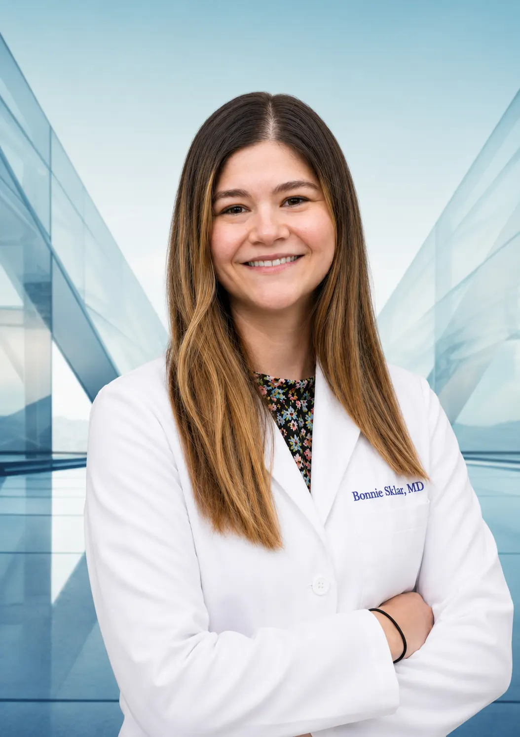
Dr. Sklar sees pterygium patients across Berg-Feinfield's Los Angeles-area offices, with surgical consultations available throughout the region.
2625 W. Alameda Ave.
Suite 208
Burbank, CA 91505
(818) 845-3557
13320 Riverside Drive
Suite 114
Sherman Oaks, CA 91423
(818) 501-3937
462 N. Linden Drive
Suite 441
Beverly Hills, CA 90212
(866) 273-3327
638 W. Duarte Road
Suite 10
Arcadia, CA 91007
(626) 795-9793
27335 Tourney Road
Suite 210
Valencia, CA 91355
(866) 273-3327
Whether your pterygium is small and just getting started or has been growing for years, Dr. Sklar and the Berg-Feinfield cornea team will give you an honest evaluation and a clear path forward.
BERG-FEINFIELD · AFTER HOURS
Questions about your vision? Talk with our AI assistant about LASIK, cataract surgery, or general eye care.
Talk with ClaireAI assistant for general information, not medical advice or emergency care. AI can make mistakes. Confirm medical information with your eye care professional.