Diagnosis, management & surgical removal by Dr. Bonnie Sklar
Pterygium — commonly called surfer’s eye — is one of the most common eye conditions in sunny, outdoor-active cities like Los Angeles. It’s treatable, and when surgery is needed, the right technique makes all the difference in preventing it from coming back.
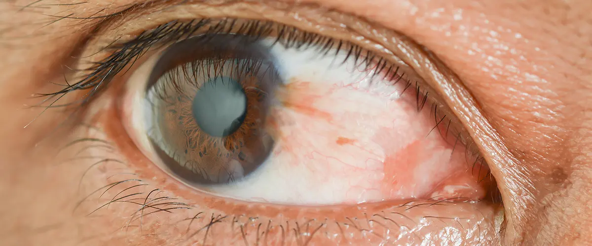
A pterygium (pronounced teh-RIJ-ee-um) is a non-cancerous growth of fibrovascular tissue that begins on the conjunctiva — the clear membrane covering the white of your eye — and spreads onto the surface of the cornea. It typically starts on the nasal side of the eye and grows horizontally toward the pupil. In some cases, both eyes are affected.
The tissue is wedge-shaped and may appear pink, fleshy, or slightly opaque. Small pterygia often cause nothing more than mild redness and irritation. Larger ones can induce corneal astigmatism, distort vision, and — in advanced cases — cover the visual axis and significantly impair sight.
Despite the alarming appearance, a pterygium is not cancerous and does not spread to other parts of the body. However, it will not go away on its own, and once it begins growing toward the center of the cornea, monitoring and eventual surgical removal is typically recommended.
Los Angeles sits between the 33rd and 34th parallels — a UV-intense latitude with year-round sun, low humidity, persistent Santa Ana winds, and a population that spends an enormous amount of time outdoors. Surfing, hiking, cycling, beach volleyball, and open-air work all contribute to cumulative UV and wind exposure. This combination makes pterygium significantly more prevalent in LA than in less sun-exposed cities.
Pterygium symptoms range from mildly bothersome to vision-impairing depending on size and location. Common signs include:
If your pterygium is growing, causing significant discomfort, distorting your vision, or approaching the pupil — see a cornea specialist. The longer a pterygium is allowed to grow onto the cornea, the more corneal tissue is involved, and the more complex the surgery becomes. Earlier is better.
Pterygia are graded by how far they extend onto the cornea. Your grade determines the urgency of treatment and influences the surgical approach if removal is needed.
The pterygium reaches the edge of the cornea (limbus) but has not yet grown onto the corneal surface. Little or no corneal involvement. Usually asymptomatic beyond mild redness.
MonitorThe pterygium extends onto the cornea but has not reached the pupil margin. May cause mild astigmatism and foreign body sensation. Conservative management or surgery depending on growth rate.
Manage or Consider SurgeryThe pterygium reaches the edge of the pupil. Significant induced astigmatism and visual distortion are common. Surgical removal is typically recommended at this stage.
Surgery RecommendedThe pterygium crosses the pupil and obscures the visual axis. Significant vision loss. Surgical removal is urgent, though corneal scarring beneath the growth may affect the final visual outcome.
Surgery UrgentTreatment depends on grade, growth rate, and symptoms. Many small pterygia are monitored for years. When growth, vision impact, or comfort cross a threshold, surgical removal is the only definitive treatment.
Artificial tears and lubricating drops reduce dryness, irritation, and the foreign body sensation associated with pterygium. They don’t stop growth but significantly improve comfort and are appropriate for mild, non-progressive pterygia.
Wraparound sunglasses with UV400 protection, wide-brimmed hats, and reducing peak-hour sun exposure are the most important steps in slowing pterygium growth and preventing recurrence after surgery. This is especially critical in Los Angeles, where UV exposure is year-round and cumulative.
When a pterygium becomes acutely inflamed — red, irritated, and uncomfortable — a short course of mild topical steroid or non-steroidal anti-inflammatory drops can reduce the inflammation. This is a temporary measure and does not treat the underlying growth.
The only way to remove a pterygium permanently is surgical excision. Dr. Sklar performs pterygium surgery using the conjunctival autograft technique — the current gold standard — which has a significantly lower recurrence rate than older approaches.
The conjunctival autograft technique involves removing the pterygium and replacing the bare area with a small graft of healthy tissue taken from the patient’s own conjunctiva — typically from the upper part of the same eye, where it is hidden under the eyelid.
Pterygium is largely a UV-related condition. In Los Angeles, year-round sun and an outdoor lifestyle mean cumulative exposure adds up quickly. These habits meaningfully reduce both first-time occurrence and post-surgical recurrence.

The single most important prevention measure. Wraparound styles block UV from the side as well as the front. Look for UV400 certification, which blocks 99–100% of UV-A and UV-B rays.
A hat with a 3-inch or wider brim reduces UV exposure to the eyes by up to 50% on a clear day. Especially important for outdoor workers, hikers, surfers, and cyclists in the LA area.
UV intensity is highest between 10am and 2pm. Scheduling outdoor activities for early morning or late afternoon reduces cumulative exposure significantly over a lifetime.
Wind and dry air — especially during Santa Ana conditions — contribute to pterygium formation and irritation. Regular use of preservative-free artificial tears reduces exposure-related irritation.
Annual comprehensive exams allow early detection of pterygium growth before it becomes symptomatic or reaches the corneal visual axis — keeping your treatment options simple.
Outdoor workers, construction professionals, landscapers, and anyone regularly exposed to dust, debris, or reflected UV should wear protective eyewear specifically designed for occupational use.
Dr. Bonnie Sklar is Berg-Feinfield’s fellowship-trained cornea specialist and performs pterygium excision with conjunctival autograft for patients throughout the Los Angeles area. Her fellowship at Duke University Eye Center — one of the nation’s most respected cornea programs — included extensive training in anterior segment surgery and ocular surface disease.
In addition to pterygium surgery, Dr. Sklar’s practice covers the full spectrum of corneal disease: keratoconus and cross-linking, Fuchs’ dystrophy, corneal transplantation (DMEK, DSEK, PKP), complex cataract surgery, and ocular surface disease. Patients referred for pterygium evaluation benefit from access to a full-scope corneal specialist, not a generalist.
View Dr. Sklar’s full profile →Not every pterygium needs to be removed immediately. The decision to proceed with surgery is based on a combination of factors — not just the size of the growth. Dr. Sklar will evaluate your specific situation and give you an honest recommendation.
Surgery is typically recommended when one or more of the following apply:
For referring optometrists: Berg-Feinfield welcomes pterygium surgical referrals. Co-management is available at all five locations. Dr. Sklar will communicate findings and post-op instructions back to your office.
OD referral information →Dr. Sklar sees pterygium patients at Berg-Feinfield’s Burbank, Sherman Oaks, Beverly Hills, and Valencia offices. Surgical consultations available across the Los Angeles area.
Berg-Feinfield provides pterygium evaluation and surgical removal to patients throughout Los Angeles County. Communities served include Burbank, Glendale, Sherman Oaks, Encino, Studio City, North Hollywood, Van Nuys, Tarzana, Beverly Hills, West Hollywood, Bel Air, Brentwood, Santa Monica, Malibu, Culver City, Arcadia, Pasadena, Monrovia, Temple City, Valencia, Stevenson Ranch, Santa Clarita, Saugus, Newhall, Canyon Country, and surrounding communities throughout the greater Los Angeles metropolitan area.
A pterygium is not cancerous and does not spread to other parts of the body. However, it should be evaluated by an eye care professional to confirm the diagnosis — a small number of conditions that can look like a pterygium are more serious. Once confirmed as a pterygium, regular monitoring is important because continued growth onto the cornea can impair vision and make surgical removal more complex. The earlier a growing pterygium is addressed, the simpler the surgery and the better the outcome.
Recurrence is possible with any pterygium surgery technique. The conjunctival autograft method significantly reduces recurrence rates compared to older bare-sclera excision. Post-surgical UV protection — sunglasses, hats, and reducing peak-hour sun exposure — is the single most important factor in keeping a pterygium from coming back. Younger patients and those with a history of heavy sun exposure have higher recurrence risk. Dr. Sklar will discuss your individual risk level and post-operative care at your consultation.
Most patients return to normal daily activities within 1–2 weeks. The eye is typically red and mildly uncomfortable for the first several days. Anti-inflammatory and lubricating drops are used during healing. You should plan to avoid swimming, dusty environments, and contact lens wear for several weeks as directed. Driving is generally possible within a day or two for most patients, but you will need someone to drive you home on the day of surgery. Final visual stabilization may take a few months if the pterygium had induced astigmatism.
A pterygium must be removed and fully healed before LASIK or any other corneal refractive surgery can be performed. The pterygium distorts the corneal surface and induces irregular astigmatism that would make LASIK measurements inaccurate. Additionally, the UV light used in LASIK can stimulate pterygium growth. The recommended sequence is: remove the pterygium, wait for complete healing and corneal stabilization (typically 6–12 months), then proceed with LASIK if you are otherwise a good candidate.
Pterygium surgery is generally covered by medical insurance when it is medically necessary — which is typically the case when the pterygium is causing vision problems, significant discomfort, or documented growth. Coverage may be more limited for purely cosmetic removal of a small, asymptomatic pterygium. Our team will verify your benefits and obtain prior authorization before scheduling your procedure. CareCredit financing is also available.
A pinguecula is a yellowish, slightly raised deposit on the conjunctiva (white of the eye) that does NOT extend onto the cornea. It is caused by the same UV and environmental factors as pterygium and is extremely common in sunny climates like Los Angeles. A pinguecula can sometimes evolve into a pterygium over time. Unlike pterygia, pingueculae rarely require surgical removal — they are managed with lubricating drops, UV protection, and monitoring. If you are unsure which you have, an evaluation will provide a clear answer.
Whether your pterygium is small and just getting started or has been growing for years, Dr. Sklar and the Berg-Feinfield cornea team will give you an honest evaluation and a clear path forward.
Call 866-2-SEE-FAR | Burbank · Sherman Oaks · Beverly Hills · Arcadia · Valencia
