Diagnosis, medical management & DMEK surgery by Dr. Bonnie Sklar
Fuchs’ dystrophy is a hereditary condition that gradually destroys the cells your cornea needs to stay clear. Left untreated, it leads to permanent vision loss — but caught early, it can be managed. When surgery is needed, DMEK offers excellent outcomes with low rejection risk and fast recovery.

The cornea — the clear front surface of your eye — depends on a single layer of cells on its inner surface to stay transparent. These cells, called corneal endothelial cells, act as pumps that continuously remove excess fluid from the cornea. When they work properly, the cornea stays clear. When they don’t, it swells with fluid and clouds your vision.
Fuchs’ endothelial dystrophy is a hereditary condition in which these cells gradually deteriorate and die over time. As the cell count drops, the cornea loses its ability to regulate fluid and begins to swell — a process called corneal edema. The result is increasingly blurry, foggy, or hazy vision that gets worse as the disease progresses.
Fuchs’ dystrophy is more common than most people realize. It affects roughly 4% of people over age 40, is more prevalent in women than men, and tends to run in families. Many people are unaware they have it until vision changes become difficult to ignore — often in their 50s or 60s.
During sleep, the eyes are closed and cannot evaporate excess fluid. As a result, the cornea is most swollen — and vision most blurry — immediately after waking. As the day goes on and the eye is open to air, some fluid evaporates and vision temporarily improves. This pattern of morning blur that clears as the day progresses is one of the earliest and most telling signs of Fuchs’ dystrophy.
Fuchs’ dystrophy symptoms typically begin gradually and worsen over years. Early-stage disease may cause only mild visual disturbances. Advanced disease significantly impairs vision. Common signs include:
If you have a family history of Fuchs’ dystrophy: Regular corneal evaluations are recommended even before symptoms appear. Early detection significantly expands your treatment options and timeline.
Fuchs’ dystrophy progresses through distinct stages over years or decades. Understanding your current stage is essential to determining the right treatment approach — and the right timing.

Tiny deposits called guttae appear on the corneal endothelium — visible on slit lamp exam. Endothelial cell density begins to decline. Vision may be normal or only mildly affected. Most patients are unaware they have Fuchs’ at this stage.
Management focuses on monitoring and symptom relief. Hypertonic saline drops can help draw excess fluid from the cornea and ease morning blur.
Manage & MonitorThe cornea begins retaining fluid in the stroma (middle layer). Vision becomes noticeably blurry — particularly in the morning — and glare and halos worsen. Corneal thickness increases measurably on pachymetry.
Hypertonic drops and a hair dryer (held at arm’s length to gently dry the corneal surface) can provide temporary relief. Surgical planning often begins at this stage.
Monitor for SurgeryFluid breaks through to the epithelium (outer surface), forming painful blisters called bullae. Vision is severely impaired throughout the day. Significant pain and discomfort accompany visual loss. Scarring may also begin to develop.
Corneal transplant surgery — specifically DMEK — is typically required to restore vision and relieve pain at this stage.
Surgery IndicatedTreatment depends entirely on what stage your disease has reached. Early Fuchs’ is managed medically. Advanced disease is treated surgically — and today’s surgical options are dramatically better than what was available even a decade ago.
DMEK represents the most significant advance in Fuchs’ dystrophy treatment in decades. Because only the diseased inner layer is replaced — not the entire cornea — the procedure offers substantial advantages over older approaches.
For patients with Fuchs’ dystrophy who have been told they need a corneal transplant, DMEK represents the current standard of care — and the difference in recovery compared to full-thickness PKP is significant.
Full DMEK Guide →Diagnosing Fuchs’ dystrophy and staging it accurately requires more than a standard eye exam. Dr. Sklar uses a combination of advanced imaging and clinical tests to build a complete picture of your corneal health.
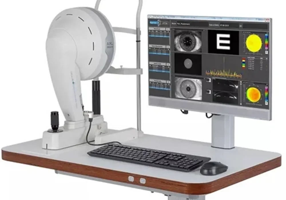
The primary tool for detecting guttae — the tiny corneal deposits that are the hallmark of Fuchs’ dystrophy. A trained cornea specialist can detect early guttae before any symptoms occur.
Photographs and counts the corneal endothelial cells, giving a precise cell density measurement. Declining cell counts are a key marker of disease progression and help guide timing of surgery.
Measures corneal thickness at multiple points. A cornea that is swelling with fluid will be measurably thicker than normal — pachymetry quantifies this and tracks change over time.
3D mapping of the corneal surface and internal structure. Used to detect irregular astigmatism caused by corneal swelling and to guide surgical planning for DMEK.
Dr. Bonnie Sklar is Berg-Feinfield’s fellowship-trained cornea specialist, with particular expertise in the surgical management of Fuchs’ endothelial dystrophy. She completed her cornea fellowship at Duke University Eye Center — one of the nation’s most respected programs — and her ophthalmology residency at Wills Eye Hospital in Philadelphia.
Her clinical practice centers on the full spectrum of corneal transplantation including DMEK, DSEK, and PKP, as well as keratoconus management, cross-linking, and ocular surface disease. For patients with Fuchs’ dystrophy considering DMEK, Dr. Sklar’s fellowship training gives her the specialized background this technically demanding procedure requires.
View Dr. Sklar’s full profile →Many patients with Fuchs’ dystrophy are first identified by their optometrist — either during a routine exam or when investigating vision changes that glasses can’t fix. If your OD has mentioned guttae, corneal swelling, or Fuchs’ dystrophy, a referral to a cornea specialist is the appropriate next step.
You don’t need to be symptomatic to benefit from a cornea specialist evaluation. Early staging — even in the absence of significant symptoms — helps establish a baseline and allows for properly timed intervention if the disease progresses.
A cornea specialist evaluation is particularly important if:
For referring optometrists: Berg-Feinfield welcomes co-management referrals for Fuchs’ dystrophy evaluation, surgical consultation, and post-operative care at all five locations.
OD referral information →Dr. Sklar sees Fuchs’ dystrophy patients at Berg-Feinfield’s Burbank, Sherman Oaks, Beverly Hills, and Valencia offices. Comprehensive corneal evaluations and DMEK surgical consultations are available across the Los Angeles area.
Berg-Feinfield provides Fuchs’ dystrophy diagnosis, medical management, and DMEK surgical treatment to patients throughout Los Angeles County. Communities served include Burbank, Glendale, Sherman Oaks, Encino, Studio City, North Hollywood, Van Nuys, Tarzana, Beverly Hills, West Hollywood, Bel Air, Brentwood, Santa Monica, Culver City, Arcadia, Pasadena, Monrovia, Temple City, Valencia, Stevenson Ranch, Santa Clarita, and surrounding communities throughout the greater Los Angeles metropolitan area.
Yes. Fuchs’ dystrophy has a strong hereditary component and tends to run in families. It is associated with mutations in genes that affect corneal endothelial cell function. If a parent has Fuchs’ dystrophy, their children have a roughly 50% chance of inheriting the condition. Regular eye exams — including slit lamp evaluation of the cornea — are recommended for first-degree relatives of anyone diagnosed with Fuchs’.
No. Many people with Fuchs’ dystrophy have early-stage disease that remains manageable with hypertonic saline drops and routine monitoring for years or even decades. Surgery becomes appropriate when vision loss significantly affects daily function, when corneal swelling progresses to the point where drops no longer provide adequate relief, or when painful surface blistering (bullous keratopathy) develops. The decision to proceed with DMEK is individualized — it depends on disease stage, visual demands, and your overall health.
Yes, but it requires careful planning. Cataract surgery stresses corneal endothelial cells, and in patients with Fuchs’ dystrophy, the reduced cell reserve means the cornea may decompensate after an otherwise routine procedure. In patients with moderate to severe Fuchs’, combining cataract surgery with DMEK in a single procedure (triple procedure) or staging them sequentially is often the preferred approach. A thorough corneal evaluation — including specular microscopy and pachymetry — before any intraocular surgery is essential. Dr. Sklar evaluates all such cases individually.
DMEK is performed as an outpatient procedure and typically takes 1–2 hours. After surgery, patients are required to maintain face-down positioning for 24 hours to allow the thin donor graft to attach properly. Most patients notice meaningful visual improvement within weeks. Vision continues to refine over several months. Long-term anti-rejection steroid eye drops are required. Compared to older full-thickness PKP transplantation, DMEK offers dramatically faster recovery, lower rejection risk, and better visual outcomes for most Fuchs’ patients.
DMEK is covered by most medical insurance plans including Medicare when it is medically necessary — which it typically is for advanced Fuchs’ dystrophy causing significant vision impairment. Coverage includes the surgeon’s fee, facility fee, anesthesia, and donor tissue. Prior authorization is generally required. Berg-Feinfield’s team verifies your benefits and manages the authorization process before scheduling your procedure.
This is actually a good situation to be in — early detection gives you the most options. If guttae have been identified but you have no significant symptoms, a baseline evaluation with a cornea specialist is the right next step. Dr. Sklar will perform specular microscopy to quantify your endothelial cell count, pachymetry to measure corneal thickness, and corneal imaging to document the extent of guttae. This baseline data is invaluable for tracking progression over time and determining if and when intervention is appropriate. Many patients with early Fuchs’ need nothing more than annual monitoring for years.
Whether you’ve just been told you have guttae or you’ve been living with Fuchs’ dystrophy for years, Dr. Sklar and the Berg-Feinfield cornea team will give you an honest assessment of where you are and what your options are.
Call 866-2-SEE-FAR | Burbank · Sherman Oaks · Beverly Hills · Arcadia · Valencia
